

Dental Fluorosis
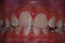
Figure 1. Very mild DF
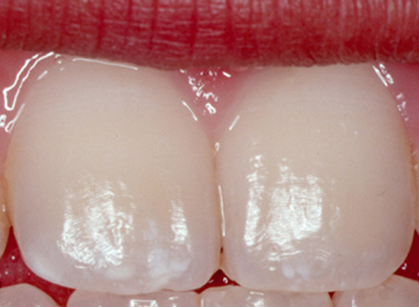
Source: Centers for Disease Control and Prevention
Dental fluorosis (DF) results from the ingestion of excessive amounts of fluoride, from all sources, during tooth development. This results in dysmineralization of the enamel. Very mild/mild DF presents as barely discernable white striae, and mottled areas of enamel.1Denbesten P, Li W. Chronic fluoride toxicity: dental fluorosis. Monogr Oral Sci 2011;22:81-96.,2Fejerskov O, Kidd E. Dental Caries - The disease and its clinical management. 2nd ed. USA: Wiley-Blackwell, 2008 These can be a cosmetic problem. (Figure 1) In contrast, severe DF results in pitted and malformed areas of enamel with porosities, brittle enamel, and altered dental morphology.1Denbesten P, Li W. Chronic fluoride toxicity: dental fluorosis. Monogr Oral Sci 2011;22:81-96.,2Fejerskov O, Kidd E. Dental Caries - The disease and its clinical management. 2nd ed. USA: Wiley-Blackwell, 2008
The risk of DF and its severity depend on the total amount of fluoride ingested, the duration of time for which it was ingested and age of the child (i.e., the stage of tooth development).3Fluoride Recommendations Work Group. Recommendations for using fluoride to prevent and control dental caries in the United States. MMWR Recomm Rep 2001;50(RR-14):1-42. In order for DF to develop, excess fluoride must be ingested prior to pre-eruptive enamel maturation. DF cannot develop after this phase. Crowns in the permanent dentition are fully formed by age 3 for the first permanent molars, and by 4 to 5 years-of-age the permanent central and lateral incisor crowns are fully formed.4American Association of Pediatric Dentists. Dental growth and development. Reference Manual V30/N6/18/19. Available at: https://www.aapd.org/globalassets/media/policies_guidelines/r_dentalgrowth.pdf. By age 8, crowns are fully formed for all of the permanent dentition, except for third molars.4American Association of Pediatric Dentists. Dental growth and development. Reference Manual V30/N6/18/19. Available at: https://www.aapd.org/globalassets/media/policies_guidelines/r_dentalgrowth.pdf. (Table 1)
| Table 1. Age for fully formed crowns for permanent teeth except third molars4American Association of Pediatric Dentists. Dental growth and development. Reference Manual V30/N6/18/19. Available at: https://www.aapd.org/globalassets/media/policies_guidelines/r_dentalgrowth.pdf. | |
|---|---|
| First permanent molars | By age 3 years |
| Central and lateral incisors | By age 4 to 5 years |
| First premolars | By age 5 to 6 years |
| Canines, second premolars | By age 6 to 7 years |
| Second molars | By age 7 to 8 years |
Prevalence of DF
Figure 2. Prevalence of DF in the United States, 1999-2004
In the United States, DF is generally very mild or mild.5Beltrán-Aguilar ED, Barker Lk, Canto MT, Dye BA, Gooch BF, et al. Surveillance for Dental Caries, Dental Sealants, Tooth Retention, Edentulism, and Enamel Fluorosis --- United States, 1988--1994 and 1999--2002. MMWR 2005;54(03):1-44. Based on the National Health and Nutrition Examination Survey (NHANES), 1999–2004, 40.7% of adolescents 12 to 15 years-of-age experienced DF.5Beltrán-Aguilar ED, Barker Lk, Canto MT, Dye BA, Gooch BF, et al. Surveillance for Dental Caries, Dental Sealants, Tooth Retention, Edentulism, and Enamel Fluorosis --- United States, 1988--1994 and 1999--2002. MMWR 2005;54(03):1-44. Very mild and mild DF was present in 28.5% and 8.6% of adolescents, respectively, and moderate to severe DF in 3.6% of individuals. (Figure 2) A prevalence of less than 1% was found for severe DF.6Beltrán-Aguilar ED, Barker L, Dye BA. Prevalence and severity of dental fluorosis in the United States, 1999-2004. NCHS Data Brief 2010;(53):1-8.
In comparing a survey conducted in the 1980s with NHANES, 1999–2004, it was found that 22.6% of 12 to 15-year-olds were affected in the 1980s compared to 40.7% in 1999-2004.5Beltrán-Aguilar ED, Barker Lk, Canto MT, Dye BA, Gooch BF, et al. Surveillance for Dental Caries, Dental Sealants, Tooth Retention, Edentulism, and Enamel Fluorosis --- United States, 1988--1994 and 1999--2002. MMWR 2005;54(03):1-44. This increased prevalence was attributed to an increasing number of sources of fluoride that may be ingested. Fluoride sources that may contribute to excessive ingestion of fluoride in total include fluoridated water (intentionally or naturally fluoridated), fluoride supplements, repeated unintentional ingestion of topical fluorides, foods and drinks containing high levels of fluoride, infant formula and medications.7U.S. Department of Health and Human Services Federal Panel on Community Water Fluoridation. U.S. Public Health Service Recommendation for Fluoride Concentration in Drinking Water for the Prevention of Dental Caries. Public Health Reports July–August 2015;130:318-31.,8Levy SM, Broffitt B, Marshall TA, Eichenberger-Gilmore JM, Warren JJ. Associations between fluorosis of permanent incisors and fluoride intake from infant formula, other dietary sources and dentifrice during early childhood. J Am Dent Assoc 2010;141:1190–201. (Table 2)
| Table 2. Sources of fluoride |
|---|
| Fluoridated water (intentionally or naturally) |
| Foods and drinks containing high levels of fluoride |
| Fluoride supplements |
| Repeated unintentional ingestion of topical fluorides |
| Infant formula |
| Certain medications |
Severe DF is endemic in areas where drinking water is obtained from wells with ground water containing high concentrations of fluoride, including in the East African Rift Valley, parts of the Middle East, Indian Subcontinent, Asia, and some areas of the Americas.9Demelash H, Beyene A, Abebe Z, Melese A. Fluoride concentration in ground water and prevalence of dental fluorosis in Ethiopian Rift Valley: systematic review and meta-analysis. BMC Public Health 2019;19:1298.,10Golgire G, Shetti S, Patil A, Khairnar M, Varekar A. Estimation of fluoride level in drinking water and prevalence of dental fluorosis in Vairag Village of Solapur District, Maharashtra, India: A cross sectional study. Epidemiology (Sunnyvale) 2016;6(275):2161-1165.1000275.,11Tekle-Haimanot R, Fekadu A, Bushera B, Mekonnen Y. Fluoride levels in water and endemic fluorosis in Ethiopian Rift Valley. 1st International Workshop on Fluorosis Prevention and Defluoridation of Water. Int Soc Fluoride Res 12-16. Editors: Eli Dahi & Henrik Bregnhøj. In one review of studies in the Ethiopian Rift Valley, ground water fluoride concentration ranged from 0.4 to 36 parts per million (ppm; mg/l).11Tekle-Haimanot R, Fekadu A, Bushera B, Mekonnen Y. Fluoride levels in water and endemic fluorosis in Ethiopian Rift Valley. 1st International Workshop on Fluorosis Prevention and Defluoridation of Water. Int Soc Fluoride Res 12-16. Editors: Eli Dahi & Henrik Bregnhøj. The overall prevalence of DF was 83%, and 35% for severe fluorosis.11Tekle-Haimanot R, Fekadu A, Bushera B, Mekonnen Y. Fluoride levels in water and endemic fluorosis in Ethiopian Rift Valley. 1st International Workshop on Fluorosis Prevention and Defluoridation of Water. Int Soc Fluoride Res 12-16. Editors: Eli Dahi & Henrik Bregnhøj. In a second review with 11 studies, the mean fluoride concentration in groundwater was 6.03 ppm while some samples yielded concentrations of up to 75 ppm.9Demelash H, Beyene A, Abebe Z, Melese A. Fluoride concentration in ground water and prevalence of dental fluorosis in Ethiopian Rift Valley: systematic review and meta-analysis. BMC Public Health 2019;19:1298. A prevalence of 32%, 29% and 24% was found for mild, moderate and severe DF, respectively.
Controlling Dental Caries and Minimizing the Risk of Fluorosis
Community water fluoridation (CWF) was introduced as a public health measure to help prevent dental caries in the 1940s at a recommended concentration of 0.7–1.2 ppm, depending on outdoor air temperature.7U.S. Department of Health and Human Services Federal Panel on Community Water Fluoridation. U.S. Public Health Service Recommendation for Fluoride Concentration in Drinking Water for the Prevention of Dental Caries. Public Health Reports July–August 2015;130:318-31. However, this was prior to the availability and use of other fluoride-containing products. In 2015, the United States recommendations changed to an optimized concentration of 0.7 ppm to gain the anti-caries benefit of fluoridated water while minimizing the risk of dental fluorosis.7U.S. Department of Health and Human Services Federal Panel on Community Water Fluoridation. U.S. Public Health Service Recommendation for Fluoride Concentration in Drinking Water for the Prevention of Dental Caries. Public Health Reports July–August 2015;130:318-31. Of note, completely ceasing CWF has been shown in several studies to result in increases in dental caries. In a recent systematic review, an increase in dental caries after cessation of CWF was found in 8 of 15 studies.12McLaren L, Singhal S. Does cessation of community water fluoridation lead to an increase in tooth decay? A systematic review of published studies J Epidemiol Community Health 2016;70:934-940. Further, in 3 of 7 studies finding no increase, alternative preventive measures had later been introduced for children, including fluorides and sealants. In Juneau, Alaska, 2.02 and 2.01 procedures attributable to dental caries were performed in the up to 18 years-of-age and less than 7 years-of-age groups, respectively, in 2003. In comparison, 2.35 and 2.68 similar procedures were performed, respectively, in 2012 which was 5 years after cessation of CWF.13Meyer J, Margaritis V, Mendelsohn A. Consequences of community water fluoridation cessation for Medicaid-eligible children and adolescents in Juneau, Alaska. BMC Oral Health 2018;18:215. Disadvantaged children were disproportionately affected, and inflation-adjusted treatment costs increased by up to 111%. In another study comparing dental caries in years with and without CWF, increases in dental caries and greater dental health inequity was found after cessation of CWF.14McLaren L, McNeil DA, Potestio M, Patterson S, Thawer S, Faris P, Shi C, Shwart L. Equity in children's dental caries before and after cessation of community water fluoridation: differential impact by dental insurance status and geographic material deprivation. Int J Equity Health 2016;15:24.
In areas with naturally high levels of fluoride in drinking water (well water), defluoridation to an appropriate and safe level is recommended as well as the use of drinking water from alternative sources.11Tekle-Haimanot R, Fekadu A, Bushera B, Mekonnen Y. Fluoride levels in water and endemic fluorosis in Ethiopian Rift Valley. 1st International Workshop on Fluorosis Prevention and Defluoridation of Water. Int Soc Fluoride Res 12-16. Editors: Eli Dahi & Henrik Bregnhøj. Further, in a hot climate substantially more water is consumed, particularly if it is a dry climate, substantially increasing the dose of fluoride at a given concentration.11Tekle-Haimanot R, Fekadu A, Bushera B, Mekonnen Y. Fluoride levels in water and endemic fluorosis in Ethiopian Rift Valley. 1st International Workshop on Fluorosis Prevention and Defluoridation of Water. Int Soc Fluoride Res 12-16. Editors: Eli Dahi & Henrik Bregnhøj. As a result, in the Ethiopian Rift Valley, mottled enamel has been found at fluoride concentrations of 2 ppm and severe dental fluorosis and skeletal fluorosis at >4 ppm. It has been suggested that guidelines on drinking water in such climates should be adjusted down from the World Health Organisation’s (WHO) recommended level of 1.5 ppm to no more than 0.6 ppm.11Tekle-Haimanot R, Fekadu A, Bushera B, Mekonnen Y. Fluoride levels in water and endemic fluorosis in Ethiopian Rift Valley. 1st International Workshop on Fluorosis Prevention and Defluoridation of Water. Int Soc Fluoride Res 12-16. Editors: Eli Dahi & Henrik Bregnhøj.
Fluoride supplements
Clinicians should evaluate all potential sources of fluoride and perform a caries risk assessment when considering fluoride supplements. In the United States, fluoride supplements are not recommended for children at low risk of dental caries, or for any children at a water fluoride level higher than 0.6 ppm. Table 3 contains current American Dental Association recommendations for once-daily use of fluoride supplements, which are limited to children at high risk of dental caries.15Rozier G, Adair S, Graham F, Iafolla T, Kingman A, Kohn W, et al. Evidence-based clinical recommendations on the prescription of dietary fluoride supplements for caries prevention: a report of the ADA Council on Scientific Affairs. Evidence-based clinical recommendations on the prescription of dietary fluoride supplements for caries prevention. J Am Dent Assoc 2010;141:1480-89. Fluoride supplements provided at higher doses recommended prior to 1994 partially explained the prevalence of fluorosis in individuals ingesting fluoride prior to pre-eruptive enamel maturation in that period.16Association of State and Territorial Dental Directors (ASTDD). Fluoride Supplement Policy Statement. Adopted January 28, 2013. Available at: https://www.astdd.org/docs/fluoride-supplement-policy-statement-january-28-2013.pdf. In a Cochrane review of randomized or quasi-randomized controlled trials with a minimum follow-up of 2 years, fluoride supplements were found to be associated with a 25% reduction in DMFS while their effect on the primary dentition was equivocal.17Tubert-Jeannin S, Auclair C, Amsallem E, Tramini P, Gerbaud L, Ruffieux C, Schulte AG, Koch MJ, Rège-Walther M, Ismail A. Fluoride supplements (tablets, drops, lozenges or chewing gums) for preventing dental caries in children. Cochrane Database Syst Rev 2011;(12):CD007592. No difference was found for the anti-caries efficacy of fluoride supplements compared to topical fluorides for the primary or permanent dentition.
| Table 3. U.S. recommendations on fluoride supplements (once daily dose) for children at high risk of dental caries15Rozier G, Adair S, Graham F, Iafolla T, Kingman A, Kohn W, et al. Evidence-based clinical recommendations on the prescription of dietary fluoride supplements for caries prevention: a report of the ADA Council on Scientific Affairs. Evidence-based clinical recommendations on the prescription of dietary fluoride supplements for caries prevention. J Am Dent Assoc 2010;141:1480-89. | ||||
|---|---|---|---|---|
| Fluoride concentration | Age | |||
| Up to 6 months | 6 months to 3 years | 3 years to 6 years | 6 years to 16 years | |
| <0.3 ppm | None | 0.25 mg/day | 0.50 mg/day | 1.0 mg/day |
| 0.3 ppm to 0.6 ppm | None | None | 0.25 mg/day | 0.50 mg/day |
| >0.6 ppm | None | None | None | None |
Home use topical fluorides
The current American Dental Association recommendations on the use of toothpaste in young children is that parents start brushing their children’s teeth twice-daily with a smear of fluoride toothpaste (the size of a grain of rice) from the time of eruption until age 3 years, and to then use a pea-sized amount of toothpaste for twice-daily brushing in children 3 to 6 years-of-age.18American Dental Association Council on Scientific Affairs. Fluoride toothpaste use for young children. J Am Dent Assoc 2014;145(2):190-91. Available at: https://jada.ada.org/article/S0002-8177(14)60226-9/pdf. Parents should be advised that children should spit out toothpaste-saliva slurry after brushing and should also be supervised during brushing once they are old enough to brush their own teeth. One estimate from 2010 suggested that ingested toothpaste in very young children was responsible for approximately 20% of total fluoride ingested.19Environmental Protection Agency (US). Fluoride: exposure and relative source contribution analysis. Washington: EPA, Office of Water, Health and Ecological Criteria Division; 2010. Available at: http://water.epa.gov/action/advisories/drinking /upload/fluoridereport.pdf. Fluoride rinses are not recommended for children under age 6 due to the risk of swallowing these. In addition, high-level fluoride pastes and gels are not recommended for use in children under age 6 years, although they may be prescribed if a dentist determines that this is indicated for a child at high risk of caries based on risk-benefit considerations.
Infant formula
In a review of infant formula, a potential risk of DF was found for powdered/concentrated formula reconstituted with fluoridated water as a part of total fluoride ingested.20Berg J, Gerweck C, Hujoel PP, King R, Krol DM, Kumar L, et al. Evidence-based clinical recommendations regarding fluoride intake from reconstituted infant formula and enamel fluorosis. A report of the American Dental Association Council on Scientific Affairs. J Am Dent Assoc 2011;142(1):79-87. It was noted that dental professionals can recommend ready-to-use formula or formula reconstituted using water containing low concentrations of fluoride or no fluoride to parents where this is a concern and whose infants are using infant formula.
Managing Fluorosis
Treatment for dental fluorosis has been found to improve oral health-related quality of life for individuals living in areas with endemic fluorosis.21Meireles SS, Goettems ML, Castro KS, Sampaio FC, Demarco FF. Dental fluorosis treatment can improve the individuals' OHRQoL? Results from a randomized clinical trial. Braz Dent J 2018;29(2):109-16. Further, in a survey using images of different levels of fluorosis, university students reported that mild and moderate fluorosis were unesthetic and that they would want treatment for these.22Gleber-Netto FO, Diniz IM, Mudado FA, Fraga MG, Vargas AM. Assessment of aesthetic perception of mild and moderate dental fluorosis levels among students from the Federal University of Minas Gerais-UFMG, Brazil. Oral Health Prev Dent 2011;9(4):339-45. Minimally-invasive options for the management of dental fluorosis include tooth whitening, microabrasion (plus or minus tooth whitening or use of CPP-ACP) and resin infiltration.23Romero MF, Babb CS, Delash J, Brackett WW. Minimally invasive esthetic improvement in a patient with dental fluorosis by using microabrasion and bleaching: A clinical report. J Prosthet Dent 2018;120(3):323-6.,24Donly KJ, O’Neill M, Croll TP. Enamel microabrasion: a microscopic evaluation of the “abrosion effect”. Quintessence Int 1992;23:175-9.,25Pini NIP, Sundfeld-Neto D, Aguiar FHB, Sundfeld RH, Martins LRM, Lovadino JR, Lima DANL. Enamel microabrasion: An overview of clinical and scientific considerations. World J Clin Cases 2015;3(1):34-41.,26Muñoz MA, Arana-Gordillo LA, Gomes GM, Gomes OM, Bombarda NH, Reis A, Loguercio AD. Alternative esthetic management of fluorosis and hypoplasia stains: blending effect obtained with resin infiltration techniques. J Esthet Restor Dent 2013;25:32-9. (Table 4)
| Table 4. Treatment options for DF |
|---|
| Tooth whitening |
| Enamel microabrasion (+/- whitening/use of CPP-ACP) |
| Resin infiltration |
| Veneers (or after tooth whitening) or crowns for more severe DF |
Tooth whitening is suitable for the treatment of mild to moderate fluorosis, and masks the white striae/mottling associated with dental fluorosis by whitening the surrounding tooth structure such that the areas blend in. Enamel microabrasion is achieved using a commercial paste containing a low concentration of hydrochloric acid and an abrasive, or phosphoric acid and pumice, that is repeatedly applied with pressure using a rubber cup and low-speed handpiece.24Donly KJ, O’Neill M, Croll TP. Enamel microabrasion: a microscopic evaluation of the “abrosion effect”. Quintessence Int 1992;23:175-9.,25Pini NIP, Sundfeld-Neto D, Aguiar FHB, Sundfeld RH, Martins LRM, Lovadino JR, Lima DANL. Enamel microabrasion: An overview of clinical and scientific considerations. World J Clin Cases 2015;3(1):34-41. This removes fluorotic/dysmineralized enamel and stains, and creates a dense, glasslike surface that alters the reflection and refraction of light and helps to mask the underlying area.24Donly KJ, O’Neill M, Croll TP. Enamel microabrasion: a microscopic evaluation of the “abrosion effect”. Quintessence Int 1992;23:175-9. (Figure 3) One study found improvements in esthetics and oral health-related quality of life one month after treatment of fluorotic areas with enamel microabrasion with or without subsequent toothwhitening.21Meireles SS, Goettems ML, Castro KS, Sampaio FC, Demarco FF. Dental fluorosis treatment can improve the individuals' OHRQoL? Results from a randomized clinical trial. Braz Dent J 2018;29(2):109-16.
Figure 3. Treatment of dysmineralized enamel with microabrasion
-
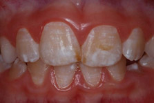
- Initial presentation
-
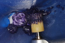
- During enamel microabrasion
-
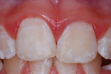
- Immediately after enamel microabrasion
-
- After enamel microabrasion
Images courtesy of Dr. Ted Croll
Resin infiltration can be performed to treat nonpitted fluorotic enamel. After isolating the teeth, the surface areas affected are first etched after which a low-viscosity resin is introduced into the enamel.26Muñoz MA, Arana-Gordillo LA, Gomes GM, Gomes OM, Bombarda NH, Reis A, Loguercio AD. Alternative esthetic management of fluorosis and hypoplasia stains: blending effect obtained with resin infiltration techniques. J Esthet Restor Dent 2013;25:32-9. The resin is then cured, resulting in the affected areas becoming denser which in turn alters how light interacts with the tooth surface and structure. This causes it to appear to be less white and to blend with adjacent tooth structure. In one study, it was found that increasing the etching time and infiltration time resulted in greater improvements in esthetics following treatment of non-pitted fluorotic white areas and stains.27Gugnani N, Pandit IK, Gupta M, Gugnani S, Soni S, Goyal V. Comparative evaluation of esthetic changes in nonpitted fluorosis stains when treated with resin infiltration, in-office bleaching, and combination therapies. J Esthet Restor Dent 2017;29(5):317-24. For patients with severe fluorosis, tooth whitening can be performed prior to placement of veneers.28Slaska B, Liebman AI, Kukleris D. Restoration of fluorosis stained teeth: A case study. Dent Clin North Am 2015;59(3):583-91. Alternatively, crowns may be placed.
Conclusions
DF is the consequence of excessive ingestion of fluoride prior to the pre-eruptive maturation during tooth development. In the United States, DF is typically very mild/mild, while in areas with naturally high concentrations of fluoride in drinking water DF can be severe. For individuals who already have DF, a number of minimally invasive treatment options is available to improve esthetics. Current recommendations on CWF, and the use of topical fluorides and supplements, are designed to optimize the proven caries-preventive benefits of fluoride while minimizing the risk of DF. It is also recommended that fluoride concentration be intentionally reduced in geographic areas with endemic fluorosis and that alternative drinking water be used. Further, that the recommended concentration in hot, dry climate be reduced below the current WHO recommendations.
It is essential that dental professionals educate parents and caregivers on the benefits of fluoride and provide advice and recommendations based on an assessment of the child’s caries risk and total fluoride exposure.
References
- 1.Denbesten P, Li W. Chronic fluoride toxicity: dental fluorosis. Monogr Oral Sci 2011;22:81-96.
- 2.Fejerskov O, Kidd E. Dental Caries - The disease and its clinical management. 2nd ed. USA: Wiley-Blackwell, 2008
- 3.Fluoride Recommendations Work Group. Recommendations for using fluoride to prevent and control dental caries in the United States. MMWR Recomm Rep 2001;50(RR-14):1-42.
- 4.American Association of Pediatric Dentists. Dental growth and development. Reference Manual V30/N6/18/19. Available at: https://www.aapd.org/globalassets/media/policies_guidelines/r_dentalgrowth.pdf.
- 5.Beltrán-Aguilar ED, Barker Lk, Canto MT, Dye BA, Gooch BF, et al. Surveillance for Dental Caries, Dental Sealants, Tooth Retention, Edentulism, and Enamel Fluorosis --- United States, 1988--1994 and 1999--2002. MMWR 2005;54(03):1-44.
- 6.Beltrán-Aguilar ED, Barker L, Dye BA. Prevalence and severity of dental fluorosis in the United States, 1999-2004. NCHS Data Brief 2010;(53):1-8.
- 7.U.S. Department of Health and Human Services Federal Panel on Community Water Fluoridation. U.S. Public Health Service Recommendation for Fluoride Concentration in Drinking Water for the Prevention of Dental Caries. Public Health Reports July–August 2015;130:318-31.
- 8.Levy SM, Broffitt B, Marshall TA, Eichenberger-Gilmore JM, Warren JJ. Associations between fluorosis of permanent incisors and fluoride intake from infant formula, other dietary sources and dentifrice during early childhood. J Am Dent Assoc 2010;141:1190–201.
- 9.Demelash H, Beyene A, Abebe Z, Melese A. Fluoride concentration in ground water and prevalence of dental fluorosis in Ethiopian Rift Valley: systematic review and meta-analysis. BMC Public Health 2019;19:1298.
- 10.Golgire G, Shetti S, Patil A, Khairnar M, Varekar A. Estimation of fluoride level in drinking water and prevalence of dental fluorosis in Vairag Village of Solapur District, Maharashtra, India: A cross sectional study. Epidemiology (Sunnyvale) 2016;6(275):2161-1165.1000275.
- 11.Tekle-Haimanot R, Fekadu A, Bushera B, Mekonnen Y. Fluoride levels in water and endemic fluorosis in Ethiopian Rift Valley. 1st International Workshop on Fluorosis Prevention and Defluoridation of Water. Int Soc Fluoride Res 12-16. Editors: Eli Dahi & Henrik Bregnhøj.
- 12.McLaren L, Singhal S. Does cessation of community water fluoridation lead to an increase in tooth decay? A systematic review of published studies J Epidemiol Community Health 2016;70:934-940.
- 13.Meyer J, Margaritis V, Mendelsohn A. Consequences of community water fluoridation cessation for Medicaid-eligible children and adolescents in Juneau, Alaska. BMC Oral Health 2018;18:215.
- 14.McLaren L, McNeil DA, Potestio M, Patterson S, Thawer S, Faris P, Shi C, Shwart L. Equity in children's dental caries before and after cessation of community water fluoridation: differential impact by dental insurance status and geographic material deprivation. Int J Equity Health 2016;15:24.
- 15.Rozier G, Adair S, Graham F, Iafolla T, Kingman A, Kohn W, et al. Evidence-based clinical recommendations on the prescription of dietary fluoride supplements for caries prevention: a report of the ADA Council on Scientific Affairs. Evidence-based clinical recommendations on the prescription of dietary fluoride supplements for caries prevention. J Am Dent Assoc 2010;141:1480-89.
- 16.Association of State and Territorial Dental Directors (ASTDD). Fluoride Supplement Policy Statement. Adopted January 28, 2013. Available at: https://www.astdd.org/docs/fluoride-supplement-policy-statement-january-28-2013.pdf.
- 17.Tubert-Jeannin S, Auclair C, Amsallem E, Tramini P, Gerbaud L, Ruffieux C, Schulte AG, Koch MJ, Rège-Walther M, Ismail A. Fluoride supplements (tablets, drops, lozenges or chewing gums) for preventing dental caries in children. Cochrane Database Syst Rev 2011;(12):CD007592.
- 18.American Dental Association Council on Scientific Affairs. Fluoride toothpaste use for young children. J Am Dent Assoc 2014;145(2):190-91. Available at: https://jada.ada.org/article/S0002-8177(14)60226-9/pdf.
- 19.Environmental Protection Agency (US). Fluoride: exposure and relative source contribution analysis. Washington: EPA, Office of Water, Health and Ecological Criteria Division; 2010. Available at: http://water.epa.gov/action/advisories/drinking /upload/fluoridereport.pdf.
- 20.Berg J, Gerweck C, Hujoel PP, King R, Krol DM, Kumar L, et al. Evidence-based clinical recommendations regarding fluoride intake from reconstituted infant formula and enamel fluorosis. A report of the American Dental Association Council on Scientific Affairs. J Am Dent Assoc 2011;142(1):79-87.
- 21.Meireles SS, Goettems ML, Castro KS, Sampaio FC, Demarco FF. Dental fluorosis treatment can improve the individuals' OHRQoL? Results from a randomized clinical trial. Braz Dent J 2018;29(2):109-16.
- 22.Gleber-Netto FO, Diniz IM, Mudado FA, Fraga MG, Vargas AM. Assessment of aesthetic perception of mild and moderate dental fluorosis levels among students from the Federal University of Minas Gerais-UFMG, Brazil. Oral Health Prev Dent 2011;9(4):339-45.
- 23.Romero MF, Babb CS, Delash J, Brackett WW. Minimally invasive esthetic improvement in a patient with dental fluorosis by using microabrasion and bleaching: A clinical report. J Prosthet Dent 2018;120(3):323-6.
- 24.Donly KJ, O’Neill M, Croll TP. Enamel microabrasion: a microscopic evaluation of the “abrosion effect”. Quintessence Int 1992;23:175-9.
- 25.Pini NIP, Sundfeld-Neto D, Aguiar FHB, Sundfeld RH, Martins LRM, Lovadino JR, Lima DANL. Enamel microabrasion: An overview of clinical and scientific considerations. World J Clin Cases 2015;3(1):34-41.
- 26.Muñoz MA, Arana-Gordillo LA, Gomes GM, Gomes OM, Bombarda NH, Reis A, Loguercio AD. Alternative esthetic management of fluorosis and hypoplasia stains: blending effect obtained with resin infiltration techniques. J Esthet Restor Dent 2013;25:32-9.
- 27.Gugnani N, Pandit IK, Gupta M, Gugnani S, Soni S, Goyal V. Comparative evaluation of esthetic changes in nonpitted fluorosis stains when treated with resin infiltration, in-office bleaching, and combination therapies. J Esthet Restor Dent 2017;29(5):317-24.
- 28.Slaska B, Liebman AI, Kukleris D. Restoration of fluorosis stained teeth: A case study. Dent Clin North Am 2015;59(3):583-91.